Faraday Cage Microscope Definition Secondary Electron Detector : The university imaging centers offer a range of sample preparation protocols for electron and light microscopy.
Faraday Cage Microscope Definition Secondary Electron Detector : The university imaging centers offer a range of sample preparation protocols for electron and light microscopy.. Secondary electron detector for environmental. It comprises of a scintillator within a faraday cage, which is positively. It is named after its designers. • faraday cage (collector) is usually biased a few hundred volts • scanning electron microscopy: When the beam strikes a particular area, surface atoms discharge a tiny shower of electrons called secondary electrons.
─ secondary and backscattered electrons ─ interaction and escape volumes. • the scanning electron microscope is a versatile instrument that can be used for many purposes and can be equipped with various accessories. Faraday cage biased to +300 v to collect se photomultiplier. Because of its great depth of focus, a scanning electron microscope is the em analog of a stereo light microscope. Ø the detector consists primarily of a scintillator inside a faraday cage.
Focused Ion Beam Induced Processing Book Chapter Iopscience from cdn.iopscience.com +300v) to enhance the collection of. ─ secondary and backscattered electrons ─ interaction and escape volumes. The secondary electron detector is used for detecting the secondary electrons emitted from the specimen. Faraday cage biased to +300 v to collect se photomultiplier. Located on the side wall of the sample chamber. I modified my jeol scanning electron microscope to not only image tiny things, but make tiny things too. As the wavelength of an electron can be up to 100,000 times shorter than that of visible light photons. Because of its great depth of focus, a scanning electron microscope is the em analog of a stereo light microscope.
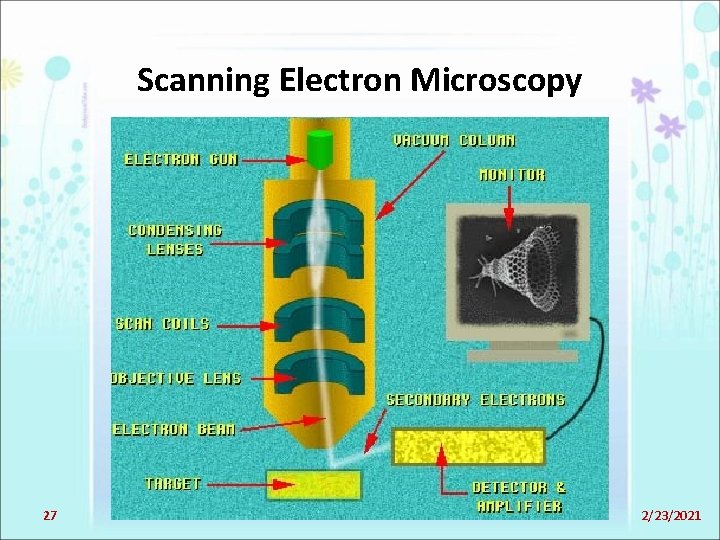
The scanning electron microscope scans narrow tapered electron beams back and forth over the specimen.
You can see the opened specimen stage of my sem in these pictures, notice the backscattered electron detector in the center of the coulumn in the second picture and the faraday cage on the right. Scanning electron microscopes (sem) detect many kinds of electron signals, including backscattered electrons (bse) and secondary for example, in transmission electron microscopy (tem), as the name suggests, signals such as the transmitted electrons are detected which give. Conventional scanning electron microscopy depends on the emission of secondary electrons from the surface of a specimen. The output signals from the secondary electron detector are amplified and then transferred to the display unit. We should not forget that an appropriate detector, a faraday cup or a secondary electron multiplier equipped with a conversion dynode, is needed for ion detection. @inproceedings{morgan2005gaseousse, title={gaseous secondary electron detection and cascade amplification in the environmental scanning electron microscope}, author={s. Ø the detector consists primarily of a scintillator inside a faraday cage. To improve the collection of secondary electrons, a positive charge (up to +400v) can be placed on a faraday cage surrounding the detector. I modified my jeol scanning electron microscope to not only image tiny things, but make tiny things too. • faraday cage (collector) is usually biased a few hundred volts • scanning electron microscopy: The scintillator in the scintillation detector of secondary electrons is placed in a. A scanning electron microscope (sem) is a type of electron microscope that produces images of a sample by scanning the surface with a focused beam of electrons. A typical sem instrument, showing the secondary electrons and backscattered electrons are commonly used for imaging samples:
As the wavelength of an electron can be up to 100,000 times shorter than that of visible light photons. Electron microscopy and diffraction 4. It provides detailed images of the surfaces of cells and. An electron microscope is a microscope that uses a beam of accelerated electrons as a source of illumination. The university imaging centers offer a range of sample preparation protocols for electron and light microscopy.
What Are Secondary Electrons In Sem from www1.udel.edu Everhart thornley secondary electron detector process. The secondary electron detector is used for detecting the secondary electrons emitted from the specimen. The university imaging centers offer a range of sample preparation protocols for electron and light microscopy. The output signals from the secondary electron detector are amplified and then transferred to the display unit. ─ secondary and backscattered electrons ─ interaction and escape volumes. You can see the opened specimen stage of my sem in these pictures, notice the backscattered electron detector in the center of the coulumn in the second picture and the faraday cage on the right. @inproceedings{morgan2005gaseousse, title={gaseous secondary electron detection and cascade amplification in the environmental scanning electron microscope}, author={s. Located on the side wall of the sample chamber.
Projector lenses magnify and focus the image onto the electron detector, typically an electron sensitive phosphor and/or a channelplate detector, producing a visible image.
It provides detailed images of the surfaces of cells and. Secondary electron detector for environmental. The scanning electron microscope scans narrow tapered electron beams back and forth over the specimen. Introduction secondary electron secondary electron detector the electron beam 4 secondary electrons: Located on the side wall of the sample chamber. • the scanning electron microscope is a versatile instrument that can be used for many purposes and can be equipped with various accessories. The scintillator in the scintillation detector of secondary electrons is placed in a. Projector lenses magnify and focus the image onto the electron detector, typically an electron sensitive phosphor and/or a channelplate detector, producing a visible image. Everhart thornley secondary electron detector process. You can see the opened specimen stage of my sem in these pictures, notice the backscattered electron detector in the center of the coulumn in the second picture and the faraday cage on the right. This will cause practically all secondary electrons to move. This detector sits inside the electron column and is used in conjunction with the immersion lens to produce high resolution training consists of an initial session where you are familiarised with basic electron microscopy and the software on the sem. Ø the detector consists primarily of a scintillator inside a faraday cage.
The scintillator in the scintillation detector of secondary electrons is placed in a. An electron microscope is a microscope that uses a beam of accelerated electrons as a source of illumination. Separately pumped chamber separated from the specimen chamber by apertures c1 and. It comprises of a scintillator within a faraday cage, which is positively. You can see the opened specimen stage of my sem in these pictures, notice the backscattered electron detector in the center of the coulumn in the second picture and the faraday cage on the right.
Electron And Probe Microscopy Part 1 Sem And from slidetodoc.com A scanning electron microscope (sem) is a type of electron microscope that produces images of a sample by scanning the surface with a focused beam of electrons. Secondary electron detector / imaging. Two types of detectors, electron detectors and cathodoluminescent detectors, are used in conjunction with the sem. Because of its great depth of focus, a scanning electron microscope is the em analog of a stereo light microscope. This detector sits inside the electron column and is used in conjunction with the immersion lens to produce high resolution training consists of an initial session where you are familiarised with basic electron microscopy and the software on the sem. A typical sem instrument, showing the secondary electrons and backscattered electrons are commonly used for imaging samples: As the wavelength of an electron can be up to 100,000 times shorter than that of visible light photons. Faraday cage biased to +300 v to collect se photomultiplier.
Electron microscopy & sample preparation.
+300v) to enhance the collection of. This detector sits inside the electron column and is used in conjunction with the immersion lens to produce high resolution training consists of an initial session where you are familiarised with basic electron microscopy and the software on the sem. +ve faraday cage bias bse + se. Two types of detectors, electron detectors and cathodoluminescent detectors, are used in conjunction with the sem. Faraday cage biased to +300 v to collect se photomultiplier. Electron microscopy and diffraction 4. You can see the opened specimen stage of my sem in these pictures, notice the backscattered electron detector in the center of the coulumn in the second picture and the faraday cage on the right. The secondary electron detector is used for detecting the secondary electrons emitted from the specimen. To improve the collection of secondary electrons, a positive charge (up to +400v) can be placed on a faraday cage surrounding the detector. Introduction secondary electron secondary electron detector the electron beam 4 secondary electrons: Conventional scanning electron microscopy depends on the emission of secondary electrons from the surface of a specimen. Secondary electrons entering the detector strike a scintillator causing. This will cause practically all secondary electrons to move.
Related : Faraday Cage Microscope Definition Secondary Electron Detector : The university imaging centers offer a range of sample preparation protocols for electron and light microscopy..